|
|
Cells: Science Education Posters & Charts
teaching resources for the science classroom and home schoolers.
|
science posters > biology > CELLS < cell biology topics
|
|
Cells are structural and functional “building block of life”. The word cell, from “cella” the Latin for small room, was adopted by English scientist Robert Hooke to describe what he saw through a microscope.
The classi cell theory was developed by Matthias Jakob Schleiden, Theodor Schwann and Rudolf Virchow.
1. All living organisms are made up of one or more cells.
2. Cells are the basic unit of life.
3. All cells arise from pre-existing cells.
4. The cell is the unit of structure, physiology, and organization in living things.
5. The cell retains a dual existence as a distinct entity and a building block in the construction of organisns.
Cell biology studies how the cell works in specialized functions like reproduction and tissue formation. Cell biologists want to know how healthy cells carry out their jobs, and how disease affects cells.
Cell biology draws heavily on genetics to research such things as human and animal cloning, how cells recognize each other during the immune response, how cancerous cells are formed, and how cells can be used to make useful products.
Cell biologists have learned how to transform fat cells into cartilage cells, which are used to replace injured tissue in joints.
|
|
|
|
|
|
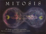
Mitosis Poster
“All change is a miracle to contemplate; but it is a miracle which is taking place every instant.” Henry David Thoreau
A: Cell membrane, B: Nucleus, C: Spindle microtubules and daughter chromosomes, D: Vacuole
|
|
|
|
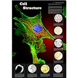
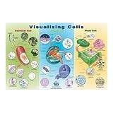
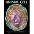
Visualizing Cells
classroom reference for a comparison of animal, plant, and bacteria cells. Includes depictions of a cell membrane, golgi apparatus, nucleus and nuclear pores, endoplasmic reticulum, chloroplasts and mitochondria.
|
|
|
|
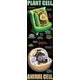
Plant and Animal Cell classroom reference illustrating animal and plant cells. Package contains a teacher's guide with background information and related activities.
|
|
|
|
|
|
|
|
|
|
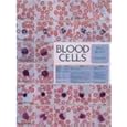
Blood Cells Chart
Every type of human blood cell is clearly illustrated, Wright-stained and magnified at 3,100X, human blood cells are vividly displayed in full-color.
• circulatory system anatomy posters
|
|
|
|
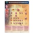
The Development of Blood Cells
Every type of human blood cell is clearly illustrated, Wright-stained and magnified at 3,100X, human blood cells are vividly displayed in full-color.
• circulatory system anatomy posters
|
|
|
|
Mitochondria, a membrane-enclosed organelle found in most eukaryotic cells, generate most of the cell's adenosine triphosphate (ATP) and play an important part in metabolic tasks.
Mitochondria have slightly different DNA and appears to be transmitted in the egg only, thus giving us important information about our evolutionary history.
The word mitochondrion comes from the Greek mitos=thread + khondrian=granule.
• Power, Sex, Suicide: Mitochondria and the Meaning of Life
|
|
|
|
Anthrax, one of the oldest recorded diseases, is caused by the bacterium Bacillus anthracis. The word anthrax is from the Greek word for coal, in reference to the black skin lesions victims develop in a cutaneous skin infection. Anthrax is highly lethal in some forms and is capable of forming long lived spores. German researcher Robert Koch isolated the Bacillus anthracis in 1877, French scientist Louis Pasteur developed the first effective vaccine for anthrax in 1881.
• Spores, Plagues and History: The Story of Anthrax
|
|
|
|
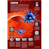
Cancer is a group of diseases in which cells grow and divide without respect to normal limits, invade and destroy adjacent tissues, and/or spread to other locations in the body (metastatize).
• The Biology of Cancer
|
|
|
|
Diatoms, one of the most common types of phytoplankton, usually consist of two asymmetrical sides with a split between them, hence the group name (Greek dia=through + temnein=to cut, i.e. cut in half).
The silica cell wall (frustule) show a wide variety of form in diatoms, some quite beautiful. Diatom fossil evidence suggests that they originated during, or before, the early Jurassic Period.
• Diatoms: Biology and Morphology of the Genera
|
|
|
|
|
|
previous page | top | cells posters > cell biology topics
|
|
I have searched the web for visual, text, and manipulative curriculum support materials - teaching posters, art prints, maps, charts, calendars, books and educational toys featuring famous people, places and events - to help teachers optimize their valuable time and budget.
Browsing the subject areas at NetPosterWorks.com is a learning experience where educators can plan context rich environments while comparing prices, special discounts, framing options and shipping from educational resources.
Thank you for starting your search for inspirational, motivational, and educational posters and learning materials at NetPosterWorks.com. If you need help please contact us.
|
|
|